What Radixact™ TomoTherapy Treats

Radixact TomoTherapy is used to treat a wide variety of cancers. In fact, Radixact TomoTherapy can treat any type of cancer that can be treated with standard radiation equipment. The advantage of Radixact Tomotherapy is that the radiation treatment is delivered with unparalled precision compared to radiation machines found in most Radiation Oncology Departments.

External-Beam Radiation Therapy

External beam radiation therapy uses a special type of very high energy X-rays to kill, fatally wound and damage cancer cells. These radiation effects shrink the tumor and help decrease the tumor’s ability to grow and spread. Radiation has its effects through direct damage to the cancer cell’s DNA, which is the cancer cell’s instruction book telling the cancer cell how to live, grow and spread.

In addition, radiation causes the formation of free radicals within the cancer cell. These free radicals cause damage to the cancer cell’s essential equipment such as proteins and cell membranes as well as add additional damage to the cell’s DNA. All of these effects of radiation taken together are what kills the cancer cells. Since radiation does not discriminate between normal cells and cancer cells, it is extremely important to limit the amount of radiation going to normal cells while delivering high does of radiation to kill and damage cancer cells. Safe and effective cancer treatment requires identifying and targeting the tumor inside the patient’s body while minimizing the amount of radiation hitting normal tissues.

At MC Radiation Oncology, we use state-of the-art technology to identify and target cancerous tumors and identify and spare normal body tissues from the radiation effects. Our treatment planning system does this by combining information about the tumor and normal tissue from PET scans, MRI scans and CT scans. This ability to combine all of these different types of scans gives us the best ability to identify and target the patient’s tumor and spare the patient’s normal tissue from the radiation effects. The Radiation Oncologist then uses all of this information to draw the tumor and the patient’s normal tissues, such as nearby organs to be avoided, into the computer treatment planning system. A treatment plan is designed in 3 dimensions to deliver the most radiation to the tumor and to minimize the radiation delivered to the normal tissues. If the tumor is in a part of the body that moves, such as the lung or in the prostate, the treatment planning includes this movement of the tumor into the treatment planning process. Once a very accurate treatment plan has been designed, the next step of the treatment process is accurately delivering the treatment to the patient. Since radiation therapy is typically delivered over a course of several treatments over several days, it is essential to compensate for subtle differences in the way the patient is positioned from treatment to treatment. At MC Radiation Oncology, we used the most advanced method for this. This method is called Image Guided Radiation Therapy (IGRT).

Image Guided Radiation Therapy using Radixact TomoTherapy

Image Guided Radiation Therapy uses an image of the part of the patient’s body where the tumor is located to guide the delivery of the radiation. This is as simple as it sounds, but at the same time is very complicated to actually do! The Radixact treatment machine used at MC Radiation Oncology creates the image used to deliver the radiation by performing a special type of CT scan immediately before each treatment. On this daily image, the tumor and the normal tissues are identified, and are then compared and matched up with the tumor and normal tissues as identified on the original treatment planning scan. The computer controlled Radixact machine performs small adjustments so that the radiation treatments are delivered as precisely as possible.

What Are the Advantages of Radixact TomoTherapy Treatment?

Treats a Wide Range of Tumors- Radixact TomoTherapy can be used to target and treat a wide variety of cancer types with very high accuracy. This includes breast cancer, prostate cancer, lung cancer, colorectal cancer, pancreatic cancer, brain cancer and many, many other types of cancer.
Customized Treatments- Since all patients are different, and all tumors are different, radiation treatments for each individual patient will all be different. At MC Radiation Oncology, each patient’s radiation treatment course is custom designed exactly for that specific patient’s tumor. Just like a fine piece of clothing, all radiation treatment programs at MC Radiation Oncology are custom tailored to the precise needs of each individual patient. Nothing comes “off the rack”! Minimized Radiation Doses- By using state-of-the Art- treatment planning systems combining PET scans, MRI scans and CT scans to identify tumors to be treated and normal tissues to be avoided and daily IGRT to confirm that the radiation plan is delivered as precisely as possible, the Radixact machine used at MC Radiation Oncology can minimize the dose the normal tissues receive and reduce to the extent possible the side-effects of treatment. This simultaneously maximizes the dose to the tumor and provides the best chance of shrinking the tumor.

Ability to Treat Multiple Tumors- Simultaneous treatment of multiple tumors is possible. This means reduced treatment time and greater patient comfort.
Non-invasive and Painless Treatment- Radiation treatment using Radixact TomoTherapy is completely non-invasive, meaning there are no needles or anything which touches the patient. The patient lies on the treatment table and relaxes while the Radixact machine does all the work! The treatments are completely painless and are essentially the same as getting a chest X-ray or a CT scan.

Benefits Include:

Leading-edge technology for individualized patient care
Radixact system enables physicians to deliver precise, non-invasive treatment while minimizing overall toxicity by minimizing radiation dose to normal tissues. This minimizes the patient’s side-effects and makes the patient’s quality of life the best it can be!
Multidisciplinary approach to treating cancer in a personalized and compassionate environment.

What Radixact™ TomoTherapy Treats

A List of the Most Common Conditions Treatable with Radixact™ TomoTherapy

A List of the Most Common Conditions Treatable with Radixact™ TomoTherapy

  • Breast Cancer
  • Prostate Cancer
  • Lung Cancer
  • Rectal Cancer
  • Pancreas Cancers
  • Head and Neck Cancer

Cancers That have Spread

  • Brain Metastasis
  • Spine Metastasis
  • Bone Metastasis

Breast

Breast Cancer

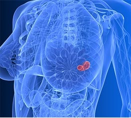
Breast cancer is very common in women. About one in nine women will get breast cancer. Chances are quite high that almost every woman has a relative with breast cancer or has a friend or acquaintance with breast cancer. Thanks largely to the use of screening mammograms (Yes, it works!) most breast cancer is caught at an early stage. This is good news, because there are excellent treatment options available. For most women with early stage breast cancer, the first decision is whether to have a mastectomy to remove the breast and check the lymph nodes or to keep her breast (breast conservation therapy). Both of these approaches offer excellent and essentially identical chances of curing the breast cancer, but are very different. After early stage breast cancer has been diagnosed, thorough research and obtaining opinions from a breast surgeon and a radiation oncologist are highly encouraged BEFORE beginning treatment.

When researching on the internet, patients should stick to “name brand” sources of information such as well-known hospitals or university medical centers. Some women with breast cancer also require chemotherapy or hormone blocking therapy. This can be incorporated with either mastectomy or breast conservation therapy and the sequencing of therapy is individualized for each unique patient.

In the “real world”, over 90% of women choose to keep the breast, that is they choose breast conservation therapy. This involves a “package deal” of surgery to remove the breast tumor, surgery (usually at the same time) to check if cancer is in the lymph nodes under the arm, and radiation treatments to the breast. The lymph nodes do not need radiation of they do not contain cancer cells. In addition, chemotherapy, “targeted therapy” and/or hormone blocking therapy may be necessary as well. The decision as to how best to combine and sequence the treatments a woman needs for her individual breast cancer is as individual as each woman! This decision is best made as part of a team approach including the breast surgeon, radiation oncologist, medical oncologist and, of course, the patient. The details of surgery and chemotherapy are best discussed with the breast surgeon and medical oncologist who will be performing those treatments. Our discussion here will focus on the radiation treatments.

The most common breast radiotherapy treatment is external beam radiation to the whole breast. This is done once a day, five days a week for a period of either 33 or 16 treatments. In the United States, the treatment approach using 33 fractions is most common. Of these 33 treatments, 28 are delivered to the entire breast and the final 5 treatments are targeted specifically to the area from which the tumor was removed. Each treatment is delivered quickly, over a period of less than 5 minutes. The patient feels nothing and there are no immediate effects during each individual treatment. Like any type of cancer therapy, radiation has both short and long term effects, and those are best discussed in detail with the radiation oncologist who will be directing the treatment.

Breast cancer therapy is one area where Radixact TomoTherapy really shines! Radixact Tomotherapy is excellent about wrapping the radiation dose around the chest to treat only the breast and greatly reduce the radiation dose to the lung and heart! This is especially true of left breast cancers since the heart is located on the left side of the chest. This greatly reduces the risk of long term radiation effects such as scarring of the lung or damage to the heart. When the lymph nodes contain cancer cells, the lymph nodes which drain lymph fluid from the breast are usually treated as well. This results in more radiation reaching the normal tissues such as the lung and the heart. However, the “collateral damage” to the lung and heart is much less with Radixact Tomotherapy than with older conventional radiation techniques such as 3D conformal radiation therapy. The cosmetic appearance of the breast after external beam radiation is excellent in most women.

Overall, external beam radiation treatment used as a part of breast conservation therapy for early stage breast cancer has a less than 5% chance of the cancer returning in the treated breast and has cure rates equal to mastectomy. These excellent results allow most women with early stage breast cancer to keep their breast with excellent cosmetic results and avoid the need for breast reconstruction or the use of breast prostheses in their bra.
A diagnosis of breast cancer is almost always life-changing. As such, many patients perform a “lifestyle reassessment” in which they re-evaluate the roles of maintaining healthy nutrition and exercise habits and well as adopting stress reducing techniques.

Many also seek out the help and advice of friends as well as to broaden their social connections with others, especially with other women who have been through the breast cancer treatment process. Some nationally recognized support groups include the Susan G Komen Foundation which specializes in breast cancer support services and its fund raising event Race for the Cure. Other national organizations which offer support for breast cancer patients as well as support to people with many other types of cancer include the American Cancer Association, Relay for Life and many many others. A Google search will quickly identify the cancer support organizations in your local area which may be able to help you during your journey.

Prostate

• Prostate cancer

TREATING PROSTATE CANCER

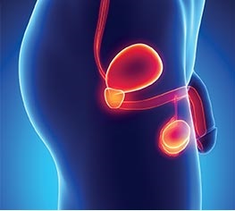
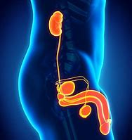
Prostate cancer is a condition in which malignant (cancer) cells occur in the prostate, a small pelvic gland which is located in the pelvis between the bladder and the rectum. Prostate cancer is a very common cancer among men. The risk factors that are definitely related to the risk of developing prostate cancer are things you can do nothing about! These are age (getting older increases the risk of prostate cancer) and having blood relatives with prostate cancer (the more blood relatives with prostate cancer and the closer they are related to you both increase the risk).
The treatment that is most appropriate for your prostate cancer depends on various factors such as your overall health status, your age, your PSA (Prostate Specific Antigen level) and the grade and stage of the prostate cancer when it is treated. And of course, your choice of the treatment you want to receive.

It is essential for you to take the time to research your treatment options, ask questions and weigh the benefits of each treatment against its potential risk of complications and side effects, as well as each treatment’s proven track record.
The most common proven treatment options for prostate cancer are: surgery (radical prostatectomy either using conventional surgical methods or robotic surgery), radiation therapy utilizing external beam or brachytherapy with or without testosterone blocking therapy and observation (watchful waiting).

External Beam Radiation therapy for prostate cancer

Radiation can be used in several different ways:
1) As a curative option, with or without hormone blocking therapy
2) If the cancer is not completely removed during surgery (prostatectomy).
3) If the tumor comes back (recurs) in the area after prostatectomy.
4) If the prostate cancer has spread causing symptoms such as pain or pressure on a normal body structure.
External beam radiation therapy is delivered with daily treatment sessions over a variable period of time with a machine called a Linear Accelerator (LINAC) which generates a beam of high energy x rays which targets the prostate cancer.
Additionally, if prostate cancer has spread into the bones, an injection of a radioactive drug (Radium-223) into the veins can help relieve pain and prolong your survival time.

External beam radiation therapy is non-invasive and painless. It is much like getting a regular X-ray, but for a longer time.

The State of the Art Radixact™ TomoTherapy linear accelerator we use at MC Radiation Oncology Center allows for greater precision in treating the prostate cancer while reducing the radiation exposure to nearby healthy tissues.
Actual treatment times using Radixact™ TomoTherapy are usually less than 5 minutes per session. These newer radiation therapy delivery techniques can offer better chances of increasing the success rate in curing prostate cancer and reducing the side effects of the radiation treatment.

Radiosurgery is another radiation related treatment option that we offer.

The most important thing to know about radiosurgery is that there is NO SURGERY involved! In radiosurgery, high-doses of radiation are delivered to the prostate gland in a few minutes in about 5 treatments. These treatments are delivered on an outpatient basis similar to conventional radiation therapy treatments. Results indicate that radiosurgery is likely as effective as surgery or conventional radiation treatments.

Other Treatment Options

Surgery : The most common curative operation for prostate cancer is called radical prostatectomy and involves removal of the entire prostate along with the adjacent glands called the seminal vesicles. This can be performed using either a conventional “open” surgical technique or using a less invasive surgical technique with a surgical robot. If a patient is considering having surgery for prostate cancer (either a conventional “open” surgery or robotic surgery) he should have a detailed consultation with the surgeon who will be doing the surgery. The patient should ask the surgeon about the possible risks of surgery and the potential side effects or complications of surgery both in the short term (days to weeks) and long term (months, years and decades) later.

Hormone Therapy: The goal of hormone therapy (also called androgen deprivation) is to lower the levels of the male hormones or androgens, such as testosterone. Prostate cancer is stimulated to grow by testosterone, thus in some cases, better results can be obtained by shutting down or drastically decreasing the body’s production of testosterone in addition to external beam radiation treatments.

Observation (Watchful Waiting, Expectant Waiting): Because some cases of prostate cancer may grow very slowly, some patients with low risk tumors can be followed using laboratory and imaging exams in a rather strict protocol with no treatment given. This approach is called watchful waiting or expectant waiting. Doctors call this expectant waiting because they expect at some point in time some prostate cancers will begin to grow more rapidly, and such cancer behavior is “expected” is some patients. When the prostate cancer changes behavior and begins to grow more rapidly, the expectant waiting approach is stopped and active treatment is started. By definition, observation involves close monitoring of the patient’s condition without giving any treatment until symptoms appear or lab values such as the PSA change.

Lung

  • Early stage Lung cancer

  • Recurring Lung cancer

  • Metastic Lung tumors

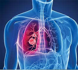
Lung cancer is the leading cause of cancer-related death in the USA. For many years, lung cancer has been the leading cause of cancer related death among men. Recently, lung cancer passed breast cancer as the leading cause of cancer related death in women as well. Early diagnosis and treatment can help. Depending on the cancer type, the cancer stage (how far advanced the cancer is) and the medical condition and the wishes of the patient various treatments could be appropriate. The possibilities include surgery, radiation therapy, chemotherapy, “targeted therapy” or any combination of these possible treatments may be recommended. Because lung cancers typically grow and spread rapidly, a “wait and watch” approach is not often recommended for lung cancer, but may be appropriate is some unusual cases.

At MC Radiation Oncology Center, we provide you with the most advanced radiation options to treat lung cancer. Lung cancer treatment is often “multimodality” or rather involves the use of several different types of medical practitioners such as surgical oncologists, medical oncologists, radiation oncologists and occasionally allied health disciplines. Our Radiation Oncologist will work in close coordination with these other medical specialists to tailor make the best treatment for each individual patient.

As noted previously, lung cancer therapy often involves the use of surgery, chemotherapy including standard chemotherapy or “targeted therapy” and radiation therapy. Any surgical procedure should be discussed with the surgeon planning to perform the operation. Any chemotherapy or “targeted therapy” should be discussed with the medical oncologist who will be performing this therapy. Likewise, any radiotherapy should be discussed with the Radiation Oncologist who will be delivering the radiotherapy.

Many factors go into deciding how radiation therapy is delivered in terms of the technique with which radiation is delivered. Radiation options for delivering radiotherapy treatments include Radiosurgery, which treats only the tumor. Radiosurgery is also called Stereotactic Body Radiation Therapy or SBRT for short. Radiation can also be delivered using conventional techniques to the tumor and lymph nodes containing the tumor. Either SBRT or conventional radiotherapy can be delivered using the most advanced radiation technology called Helical Tomotherapy using Radixact. Helical Tomotherapy using Radixact is the state of the art treating technology used at MC Radiation Oncology Center.This permits optimal targeting of the lung cancer while minimizing the radiation effects on nearby normal tissue,

SBRT, or stereotactic body radiation therapy, is also known as radiosurgery. Despite the use of the word “surgery” in its name, radiosurgery does not involve cutting into the patient to remove the tumor in an operating room using a surgical knife or anesthesia or hospitalization. Radiosurgery uses X ray of high intensity and special software that allows radiation to penetrate deep into the body while minimizing the radiation effects on normal tissues.

Advantages of TomoTherapy Radixact treatment delivery system design provides volumetric beam delivery. This customized design combined with next generation CT/PET fusion imaging provides state of the art cancer treatment for our patients.
• Shorter Treatment Time
• Non-Invasive procedure (no surgery or cutting)
• No hospital stay
• Rapid return to normal activities

Lung Cancer Facts

Each year more than 170,000 Americans are newly diagnosed with lung cancer.
• Cigarette smoking is the biggest risk factor in the development of lung cancer.
• The 5 year survival rate for lung cancer in general is less than 15% lagging far behind that of other cancers. The 5 year survival rate for early stage is approximates 70%.
• More people die from lung cancer than from breast cancer, prostate cancer, colon cancer, and pancreatic cancer combined.
• Smokers and others at high risk for lung cancer such as asbestos exposure should consult their physicians for appropriate lung cancer checks and early detection procedures.
• The time proven saying about smoking still holds true today: If you don’t smoke, don’t start smoking. If you do smoke, stop. Now.
Keep in mind statistics do not necessarily reflect the specifics of an individual patient. Cancer statistics are based on the total population and are compiled for groups with similar types of lung cancer and similar lung cancer stages, ranging early stage through late stage disease.

The two main types of lung cancer are non-small cell lung cancer and small cell lung cancer.  These names refer to the type of cell the lung cancer comes from.

Non-small cell lung cancer is the most common type of lung cancer and accounts for 84 percent of cases. There are several different types of non-small cell lung cancer, with the main types including:
• Adenocarcinoma – a cancer that forms in the outer parts of the lung, usually from the glands inside the lung.
• Squamous cell carcinoma – a cancer that forms from an abnormal cell lining the airway.
• Large cell carcinoma – a kind of non-small cell lung cancer, but the cell it starts from may not be known.
Small cell lung cancer is less common and accounts for 16 percent of cases. Although the cells are small, they multiply quickly and can form large tumors that may spread throughout the body. This type of lung cancer is especially aggressive and is often associated with heavy smoking. Small cell lung cancer is usually treated with a combination of chemotherapy and radiation therapy.

Care During Your Treatment

Cancer treatment can be difficult. You have many issues to cope with. Your oncology team, along with family and friends are available to help.
• Get plenty of rest during treatment. Drink plenty of fluids and eat a well-balanced diet. Your medical care team can provide you with may helpful suggestions to make life easier and better.
• If you are having problems, tell your doctor about your problems. The sooner the doctor knows there are problems, the sooner action can be taken to help you! The old adage that an ounce of prevention is worth a pound of cure rings true. Doctors cannot read minds! Your doctor cannot help you unless he or she knows what is going on!
• Ask if you are unsure about anything. There are no stupid questions.
• Tell your doctor about any medications, vitamins or supplements you are taking to make sure they are safe to use during radiation therapy.
• Nutrition is important. Let your doctor know if you have trouble swallowing, food tastes funny or you have trouble eating or have a poor appetite.
• Treat the skin exposed to radiation with special care. If you note any skin irritation, inform your radiation team about the irritation and ask for advice.

RECTAL

Rectal Cancer

Colorectal cancer is the 4th most common type of cancer diagnosed in the United States. Famous people with colorectal cancer include Charles Schultz, the creator or the Peanuts comic strip, Milton Berle, and Ronald Reagan. The rectum is the last part of the large bowel, and is largely responsible for reabsorbing water used in the digestive process and creating solid stool. The colon is a tube made up of a muscle layer which contracts to move the stool along, and an inner layer of glands which line this muscular tube. It is from this layer of glands that most colorectal cancers develop.

Medicine has learned that most colorectal cancers develop from polys, or small outgrowths or “tags” extending from the lining of the large bowel. Most of these polyps begin as benign (non-cancerous) lumps of cells, but over time some of these polyps become cancerous. This is the reason why screening colonoscopy is recommended to identify and remove polyps before the polyps have a chance to become cancerous. If removed completely, benign, pre-cancerous or very early stage cancerous polyps require no therapy in addition to the removal during colonoscopy. If polyps become cancerous and invade into the muscular wall of the large bowel, medical therapy is different because it usually involves surgery, and in addition it can involve chemotherapy and/or radiation treatments as well.

The exact location of the cancer within the large bowel is important since the location of the cancer largely determines the therapy which is recommended. Cancers of the first 2/3s or so of the large bowel are called colon cancer. When cancer invades the muscle wall of the colon, the portion of the colon containing the cancer is removed and the bowel is reconnected avoiding the need for a colostomy bag in most cases. Depending on other factors, such as cancer within the lymph nodes, chemotherapy may be recommended as well. Radiation is usually not recommended for colon cancers.
Cancers occurring in the last part of the colon before the anus are called rectal cancers. These cancers are usually treated with a combination of daily radiation treatments along with chemotherapy followed by surgery to remove the cancerous part of the rectum. In most cases, the bowel can be “re-connected” and the need for a colostomy bag can be avoided.

Overall, the best treatment for colorectal cancer is preventative. This involves having regular colonoscopy to remove polyps before they have the chance to become cancerous. If polyps become cancerous and are caught at an early stage, treatment is curative in the great majority of patients. If the cancer has spread, there are many research trials looking at new treatment approaches such as stereotactic body radiosurgery for isolated spread to the liver or lungs as well as exploring the use of new chemotherapy drugs.

PANCREAS

Pancreatic Cancer occurs in 55,000 people each year and kills 45,000 of them. Pancreatic cancer is a very difficult cancer to cure, even when caught at a very early stage, which is in itself uncommon because the pancreas is located in a part of the body where it rarely causes symptoms until it is advanced. The most common type of pancreatic cancer is adenocarcinoma, which arises from the glandular part of the pancreas. Well-known people diagnosed with pancreatic cancer include Alex Trebec, John Hurt, and Steve Jobs and Arethra Franklin (both of whom were diagnosed with a rare form of pancreatic cancer, neuroendocrine cancer).

Surgery to remove the tumor is the most effective treatment, but is only offered if the entire tumor can be removed, because removal of less than the entire tumor is of no benefit to the patient. When surgery is possible, it is a very major surgery called a “Whipple Procedure”. This involves removal of the pancreas, a portion of the small bowel, the gall bladder, bile duct, and sometimes a part of the stomach. Because most pancreatic cancers are diagnosed at a more advanced stage, surgery is not often an option. These patients are usually treated with a combination of chemotherapy and radiation.

Treatment of pancreatic cancer is one of the areas where helical tomotherapy using Radixact really shines! The pancreas and its lymph nodes are located in the middle of the body tucked between many very sensitive normal body organs such as the liver, bowel, kidneys and spinal cord great care must be taken to avoid these very sensitive structures while giving adequate treatment to the pancreatic tumor. Helical tomotherapy using radixact gives the Radiation Oncologist the ability to sculpt the radiation dose bending it around the sensitive areas to hit the tumor much in the same way as a master sculptor can shape stone or metal to create the 3D object he desires. Chemotherapy is usually given along with radiation mainly to increase the effectiveness of radiation, but also to treat microscopic deposits of cancer that may not be in the path of the radiation beam.

Head and Neck

  • Oral Cavity
  • OrPharynx
  • Hypopharynx
  • Larynx
  • Nasal Cavity and Paranasal Sinus

Head and Neck Cancer

Head and neck cancer is relatively rare, accounting for about 4% of all cancers diagnosed yearly in the US. Most head and neck cancers originate in the lining cells of the mouth, throat, or sinuses and are known as squamous cell carcinomas. Less commonly, cancers can originate in the salivary (spit) glands and are called adenocarcinomas. In the past, the main risk factors for developing head and neck carcinomas were smoking and drinking. This remains true today, but an association with Human Papilloma Virus (HPV) infection is rising, especially in cases of cancer of the oral cavity, oropharynx, and base of tongue. Another virus, the Epstein Barr Virus (EBV) is associated with some cases cancer developing in the sinuses. Some people fell diet plays a role, with an apparent increase in cancer risk for people eating a salty or preserved foods. Some feel that exposure to sawdust or asbestos increases the risk. Genetics apparently has a role, because people with Chinese ancestry have an increased risk for nasopharyngeal cancer.

Cancers of the head and neck area are named for the parts of the head and neck where they occur. These are the Oral Cavity, which includes the inner part of the lips, gums, floor of mouth under the tongue, the front part of the tongue, hard palate (roof of the mouth) the inside of the cheek and the retromolar trigone (behind the wisdom teeth). Then there is the pharynx, which is the beginning of the throat behind the oral cavity. The pharynx is divided into the oropharynx which includes the soft palate and the uvula (hangs down in the throat), the base of the tongue, and tonsils. The nasopharynx which is above the soft palate. The hypopharynx which is the throat walls around the base of tongue and larynx. The larynx or voice box. The paranasal sinuses where sinus infections occur and the salivary glands which are located throughout the mouth and in front of the ears. Mumps is a childhood viral infection of the parotid glands in front of the ears.

Head and neck cancers are all treated in a similar way, except for nasopharyngeal cancers which are always treated with chemotherapy and radiation. When caught early, surgery alone is often an option for most head and neck cancers. When caught early and the patient cannot have surgery or refuses surgery, then radiation alone is often an option. When cancers are caught at a more advanced stage, then a combination of chemotherapy and radiation is commonly used. For example, traditionally surgical removal of the voice box (larynx) was recommended for advanced cases of carcinomas of the voice box. However this is no longer the case. Advanced laryngeal cancers are now routinely treated with a combination of chemotherapy and radiation. Using this “combined modality approach” the majority of patients can save their voice box and have equal chances of being cured compared to those undergoing removal of the voice box then receiving radiation treatment after surgery. This results in a much improved quality of life in most patients undergoing combined modality therapy.

Genitourinary Cancers

  • Kidney
  • Bladder
  • Prostate

Kidney Cancer

Kidney cancer is a cancer which arises from the cells of the kidney. The kidney is an organ which cleans the blood of waste products and concentrates the waste products into urine. The most common type of kidney cancer is Renal Cell Carcinoma, which amounts to about 90% or more of all cancers arising from the kidney. There are about 74,000 new cases of renal cell carcinoma in the US each year, and it is slightly more common in men. The typical age at diagnosis is 50-70 years. Risk factors for renal cell carcinoma are smoking (which doubles the risk), obesity, and certain uncommon genetic conditions, and various other factors. Since the kidneys are located behind the upper abdominal area and are separated from the intestines and other abdominal organs, most cases of kidney cancer do not cause symptoms until they are relatively advanced, meaning it is large or has spread to other body areas such as the lymph nodes, lungs, liver, bone or brain. Famous people who have had rencal cell carcinoma include Raymond Burr and James “Hacksaw” Duggan of wrestling fame.

For early stage renal cell carcinoma, which is uncommon because most renal cell cancers are not detected at an early stage, the therapy is usually surgery to remove all or part of the kidney where the cancer is located (radical or partial nephrectomy). When caught at the earliest stage (Stage I) the chances of 5 year survival are about 80%. When caught at later stages, the chances of surviving the cancer go down rapidly to about 8% for stage IV tumors. Chemotherapy or “targeted” therapy often plays a major role in treating renal cell carcinoma.
Radiation is no longer commonly used for curative therapy of renal cell carcinomas. One exception is the possible use of stereotactic radiosurgery for treating some cases where the renal cell carcinoma has spread to very limited areas of the body. Radiation is commonly used in palliative treatment of cancer metastases (spots of cancer that have spread to other areas of the body) as a way to relieve symptoms such as pain or to treat spread of the renal cell carcinomas to the brain.

Ureteral Carcinoma

Ureteral carcinomas are cancers arising from the ureter, which is the tube that carries urine from the kidney to the bladder. Ureteral carcinomas are extremely rare. These cancers occur in only about 4 people in 1 million. Men are more likely to get ureteral carcinomas. Smoking increases the risk of getting ureteral carcinoma as does exposure to certain types of chemicals.
The most common type of ureteral carcinoma is transitional cell carcinoma, and this begins in the cells that line the ureter. Transitional cell carcinoma is also the most common type of cancer arising in the bladder. As such, it is not surprising that ureteral cancers are far more common in people with a history of previous bladder cancer. Ureteral cancer also greatly increases the risk of a patient developing bladder cancer, if they haven’t already done so. In the rare case when ureteral cancer is caught early, the main treatment is surgery to remove the ureter and/or kidney on the side where the cancer developed. Most commonly, ureteral cancers are detected at an advanced stage where the tumor is large or the cancer has spread. In those cases (the great majority) surgery can sometimes be used but chemotherapy or “targeted therapy” is often the most important treatment.

Bladder Cancers

Bladder cancer is the 6th most common type of cancer in the United States. About 80,000 new cases of bladder cancer are diagnosed each year and about 17,500 people die of this cancer yearly. Overall, the 5 year survival of all stages of bladder cancer together is good at about 77%.

The bladder is an organ in the pelvis made up of muscle which is lined by cells. By far the most common type of cell lining the bladder is the transitional cell. Malignancies arising from the transitional cells are called transitional cell carcinoma. Other and far less common types of cells found in the bladder, usually in unusual cases are adenomatous cells and squamous cells. For purposes of this discussion, we will stick with the treatment for the transitional cell carcinomas, which is by far the most common type of bladder cancer.

Catching a cancer at the earliest possible stage is essential in having the best chances of curing the cancer. This is very true of bladder cancer. A key factor going into the staging (determining how far along the cancer is) of bladder cancer is whether the cancer has invaded the muscle wall of the bladder and if it has invaded, how deep into the muscle wall the tumor has invaded. Cancers which have not yet invaded the muscle wall of the bladder are called “carcinoma in situ” which means pre-invasive cancer. Transitional cell carcinoma in situ of the bladder is generally treated by “scraping” the pre-cancerous tumor off the bladder wall. Usually this is followed by putting a form of chemotherapy or immunotherapy into the bladder.

If the cancer has invaded into the muscle wall of the bladder, but the tumor isn’t very large or doesn’t go all the way through the bladder wall, then a combination of chemotherapy and daily radiation treatments can often avoid the need to remove the bladder and to have a cystostomy bag. If removal of the bladder is necessary, sometimes radiation and/or chemotherapy after surgery are needed.

Treating Prostate Cancer

Prostate cancer is a condition in which malignant (cancer) cells occur in the prostate, a small pelvic gland which is located in the pelvis between the bladder and the rectum. Prostate cancer is a very common cancer among men. The risk factors that are definitely related to the risk of developing prostate cancer are things you can do nothing about! These are age (getting older increases the risk of prostate cancer) and having blood relatives with prostate cancer (the more blood relatives with prostate cancer and the closer they are related to you both increase the risk).
The treatment that is most appropriate for your prostate cancer depends on various factors such as your overall health status, your age, your PSA (Prostate Specific Antigen level) and the grade and stage of the prostate cancer when it is treated. And of course, your choice of the treatment you want to receive.

It is essential for you to take the time to research your treatment options, ask questions and weigh the benefits of each treatment against its potential risk of complications and side effects, as well as each treatment’s proven track record.
The most common proven treatment options for prostate cancer are: surgery (radical prostatectomy either using conventional surgical methods or robotic surgery), radiation therapy utilizing external beam or brachytherapy with or without testosterone blocking therapy and observation (watchful waiting).

External Beam Radiation therapy for prostate cancer

Radiation can be used in several different ways:

1) As a curative option, with or without hormone blocking therapy
2) If the cancer is not completely removed during surgery (prostatectomy).
3) If the tumor comes back (recurs) in the area after prostatectomy.
4) If the prostate cancer has spread causing symptoms such as pain or pressure on a normal body structure.
External beam radiation therapy is delivered with daily treatment sessions over a variable period of time with a machine called a Linear Accelerator (LINAC) which generates a beam of high energy x rays which targets the prostate cancer.

Additionally, if prostate cancer has spread into the bones, an injection of a radioactive drug (Radium-223) into the veins can help relieve pain and prolong your survival time.

External beam radiation therapy is non-invasive and painless. It is much like getting a regular X-ray, but for a longer time.
The State of the Art Radixact™ TomoTherapy linear accelerator we use at MC Radiation Oncology Center allows for greater precision in treating the prostate cancer while reducing the radiation exposure to nearby healthy tissues.

Actual treatment times using Radixact™ TomoTherapy are usually less than 5 minutes per session. These newer radiation therapy delivery techniques can offer better chances of increasing the success rate in curing prostate cancer and reducing the side effects of the radiation treatment.
Radiosurgery is another radiation related treatment option that we offer.

The most important thing to know about radiosurgery is that there is NO SURGERY involved! In radiosurgery, high-doses of radiation are delivered to the prostate gland in a few minutes in about 5 treatments. These treatments are delivered on an outpatient basis similar to conventional radiation therapy treatments. Results indicate that radiosurgery is likely as effective as surgery or conventional radiation treatments.

Other Treatment Options

Surgery : The most common curative operation for prostate cancer is called radical prostatectomy and involves removal of the entire prostate along with the adjacent glands called the seminal vesicles. This can be performed using either a conventional “open” surgical technique or using a less invasive surgical technique with a surgical robot. If a patient is considering having surgery for prostate cancer (either a conventional “open” surgery or robotic surgery) he should have a detailed consultation with the surgeon who will be doing the surgery. The patient should ask the surgeon about the possible risks of surgery and the potential side effects or complications of surgery both in the short term (days to weeks) and long term (months, years and decades) later.

Hormone Therapy: The goal of hormone therapy (also called androgen deprivation) is to lower the levels of the male hormones or androgens, such as testosterone. Prostate cancer is stimulated to grow by testosterone, thus in some cases, better results can be obtained by shutting down or drastically decreasing the body’s production of testosterone in addition to external beam radiation treatments.

Observation (Watchful Waiting, Expectant Waiting): Because some cases of prostate cancer may grow very slowly, some patients with low risk tumors can be followed using laboratory and imaging exams in a rather strict protocol with no treatment given. This approach is called watchful waiting or expectant waiting. Doctors call this expectant waiting because they expect at some point in time some prostate cancers will begin to grow more rapidly, and such cancer behavior is “expected” is some patients. When the prostate cancer changes behavior and begins to grow more rapidly, the expectant waiting approach is stopped and active treatment is started. By definition, observation involves close monitoring of the patient’s condition without giving any treatment until symptoms appear or lab values such as the PSA change.

Digestive Cancers

  • Esophagus
  • Stomach
  • Pancreas
  • Colorectal

Esophageal Cancer

Esophageal cancer is a relatively uncommon cancer with about 20,000 new cases diagnosed in the United States each year. Famous people who have had esophageal cancer include Humphrey Bogart, Robert Kardashian, and Richard Dawson. Esophageal cancer is a very serious type of cancer. Like most cancers, the cure rates for esophageal cancer relate to how far along the cancer is when it is diagnosed. Since most cases of esophageal cancer are diagnosed at a later stage, the cure rates are not very good. The chances of surviving esophageal cancer after 5 years from the diagnosis are about 50% if the cancer is confined to the esophagus and 25% when the cancer involves the near by lymph nodes. If the cancer has spread beyond the lymph nodes, the chance of survival drop substantially from that.
The esophagus is a muscular tube which connects the mouth to the stomach. Like most muscular structures in the body such as the intestines and the bladder, the esophagus is lined by special types of cells. The upper part of the esophagus is lined by squamous cells, which resemble roofing tiles under the microscope. Cancers arising from this type of cell are called squamous cell carcinomas. These cancers are often related to tobacco use, heavy drinking, and race amongst other factors. Over the last several years, another type of cancer called adenocarcinoma has become more common. Chronic reflux (acid gurgling from the stomach into the lower esophagus) causes the squamous cells normally found in the esophagus to transform into glandular cells normally found in the stomach or intestines. This is called Barrett’s Esophagus and is most commonly found at the bottom of the esophagus where it connects to the stomach. Sometimes these abnormal glandular cells can become a cancer. The most common risk factors for developing adenocarcinoma of the esophagus include chronic reflux, obesity, and being a white male. There are many other less common risk factors, but the ones mentioned are the most common.
Treatment of esophageal cancer depends on the stage of the cancer, meaning whether the cancer was detected early or at an advanced stage. Early stage esophageal cancer is rarely diagnosed, but can sometimes be treated with removal of the lining of the esophagus in the earliest cases. In other cases, early stage esophageal cancer may be treated with the removal of the esophagus. In the typical case where esophageal cancer involves the nearby lymph nodes, esophageal cancer is usually treated with a combination of chemotherapy and radiation with or without removal of the esophagus.

Stomach Cancer

Most cases of stomach cancer begin in the glands which make up the lining of the stomach. Cancers which arise in glands are called adenocarcinomas. There are about 200,000 cases of stomach cancer diagnosed in the US each year. Risk factors for developing stomach cancer are smoking, obesity, a diet rich in smoked pickled or salty foods, certain infections including Epstein Barr virus or H. Pylorii, and certain medical conditions such as pernicious anemia.
When detected at the earliest stage, when the cancer sits on top of the lining of the stomach, surgery to remove it is usually curative. This is rare. Most often, the cancer has invaded into the lining of the stomach or beyond. In those cases surgery to remove all or part of the stomach is often a major part of the treatment. Most cases will received a combination of radiation with chemotherapy in addition to surgery or instead of surgery. In very selected cases of very limited spread of the cancer, radiosurgery or surgery can be used against the limited spread of cancer in addition to treatment of the main cancer. When stomach cancer has spread and is causing symptoms, radiation is often used as a palliative treatment, designed to relieve symptoms such as pain.

Pancreas

Pancreatic Cancer occurs in 55,000 people each year and kills 45,000 of them. Pancreatic cancer is a very difficult cancer to cure, even when caught at a very early stage, which is in itself uncommon because the pancreas is located in a part of the body where it rarely causes symptoms until it is advanced. The most common type of pancreatic cancer is adenocarcinoma, which arises from the glandular part of the pancreas. Well-known people diagnosed with pancreatic cancer include Alex Trebec, John Hurt, and Steve Jobs and Arethra Franklin (both of whom were diagnosed with a rare form of pancreatic cancer, neuroendocrine cancer).

Surgery to remove the tumor is the most effective treatment, but is only offered if the entire tumor can be removed, because removal of less than the entire tumor is of no benefit to the patient. When surgery is possible, it is a very major surgery called a “Whipple Procedure”. This involves removal of the pancreas, a portion of the small bowel, the gall bladder, bile duct, and sometimes a part of the stomach. Because most pancreatic cancers are diagnosed at a more advanced stage, surgery is not often an option. These patients are usually treated with a combination of chemotherapy and radiation.
Treatment of pancreatic cancer is one of the areas where helical tomotherapy using Radixact really shines! The pancreas and its lymph nodes are located in the middle of the body tucked between many very sensitive normal body organs such as the liver, bowel, kidneys and spinal cord great care must be taken to avoid these very sensitive structures while giving adequate treatment to the pancreatic tumor. Helical tomotherapy using Radixact gives the Radiation Oncologist the ability to sculpt the radiation dose bending it around the sensitive areas to hit the tumor much in the same way as a master sculptor can shape stone or metal to create the 3D object he desires.

Chemotherapy is usually given along with radiation mainly to increase the effectiveness of radiation, but also to treat microscopic deposits of cancer that may not be in the path of the radiation beam.

Colorectal Cancer

Colorectal cancer is the 4th most common type of cancer diagnosed in the United States. The colon is the last part of the large bowel, and is largely responsible for reabsorbing water used in the digestive process and creating solid stool. The colon is s tube made up of a muscle layer which contracts to move the stool along, and an inner layer of glands which line this muscular tube. It is from this layer of glands that most colorectal cancers develop.
Medicine has learned that most colorectal cancers develop from polys, or small outgrowths or “tags” extending from the lining of the large bowel. Most of these polyps begin as benign (non-cancerous) lumps of cells, but over time some of these polyps become cancerous. This is the reason why screening colonoscopy is recommended to identify and remove polyps before the polyps have a chance to become cancerous. If removed completely, benign, pre-cancerous or very early stage cancerous polyps require no therapy in addition to the removal during colonoscopy. If polyps become cancerous and invade into the muscular wall of the large bowel, medical therapy is different because it usually involves surgery, and in addition it can involve chemotherapy and/or radiation treatments as well.
The exact location of the cancer within the large bowel is important since the location of the cancer largely determines the therapy which is recommended. Cancers of the first 2/3s or so of the large bowel are called colon cancer. When cancer invades the muscle wall of the colon, the portion of the colon containing the cancer is removed and the bowel is reconnected avoiding the need for a colostomy bag in most cases. Depending on other factors, such as cancer within the lymph nodes, chemotherapy may be recommended as well. Radiation is usually not recommended for colon cancers.
Cancers occurring in the last part of the rectum before the anus are usually treated with a combination of daily radiation treatments along with chemotherapy followed by surgery to remove the cancerous part of the rectum. In most cases, the bowel can be “re-connected” and the need for a colostomy bag can be avoided.
Overall, the best treatment for colorectal cancer is having regular colonoscopy to remove polyps before they have the chance to become cancerous. If polyps become cancerous and are caught at an early stage, treatment is curative in the great majority of patients. If the cancer has spread, there are many research trials looking at new treatment approaches such as stereotactic body radiosurgery for isolated spread to the liver or lungs as well as exploring the use of new chemotherapy drugs.

Central Nervous System (CNS)

  • Primary Brain Tumors
  •  Secondary Brain Tumors (Brain Metastasis)
  • Primary Spinal Cord Tumors

Primary Brain Tumors

Primary brain tumors originate from any cell type in the brain. In fact, there are over 120 different types of primary brain tumors. Tumors arising from glial cells (Gliomas) are the most common primary brain tumors affecting approximately 30 per 100,000 people in the United States. Most primary brain tumors are benign, but they can cause damage or death by invading or compressing nearby normal brain tissue. There are also malignant, or cancerous brain tumors. Primary brain tumors are treated with observation, surgery, surgery and radiation after surgery, or radiation alone. The choice of treatment depends on the type of brain tumor, the location of the brain tumor and the symptoms the tumor is causing the patient or the loss of function that may result if the tumor continues to grow. Brain tumors are as much individuals as are brain tumor patients and the treatment for each individual patient is personalized according to that patient’s circumstances. The individual treatment recommended is best decided in a team approach including the neurosurgeon, radiation oncologist, medical oncologist and often a physical therapist, a psychologist and a social worker.

Gliomas arise from the “supporting cells” that are believed to support and nourish the nerve cells which perform the functions of thinking, controlling muscle movement and other functions essential to life. Astrocytes give rise to the most common type of gliomas, the astrocytomas. Astrocytomas themselves can be classified according to grade from I-IV. Grade typically correlates with the aggressiveness of the tumor. Typically Grades 1 and 2 astrocytomas are slow growing tumors and may be present for years before they cause problems for the patient. Grade III astrocytomas are called anaplastic astrocytomas and have a much more aggressive behavior and always require treatment. Grade IV astrocytomas are also called Glioblastomas or Glioblastoma Multiforme. Glioblastomas are an extremely aggressive form of brain tumor that usually follows a rapid growth behavior and invades and destroys a large portion of the surrounding normal brain. These tumors are ideally treated with maximal safe surgery which is followed by a combination of daily radiation treatments combined with chemotherapy. New “targeted agent” therapy is showing promise as an alternative or as an addition to standard chemotherapy.

Other types of gliomas include ependymomas, which arise from the lining of the cavities within the brain which contain the cerebrospinal fluid. These include anaplastic ependymoma, myxopapillary ependymoma, and subependymoma. Another type of glioma is the oligodendroglioma and this type includes oligodendroglioma, anaplastic oligodendroglioma, and anaplastic oligoastrocytoma. These types of gliomas are quite rare and a detailed discussion is not appropriate here, but is best held between a patient with this type of diagnosis and the physicians who will be treating this patient.
Other primary brain tumor types include tumors arising from the brain coverings or meninges (meningiomas) and supporting tissues such as blood vessels (Hemangiomas) or abnormal connections between arteries and veins (Arteriovenous malformations) which could rupture and bleed, white blood cells (Lymphomas), nerves (Neurinomas), pituitary gland (Pituitary Adenomas) and pineal gland (Pinealomas or ).

Brain tumors are invasive and grow in a limited space within the skull by directly damaging (invading) adjacent parts of the brain as well as taking up space by their growth and putting pressure on all of the brain. Because of their location, treatment should often not be delayed. Occasionally, brain tumors in non-critical areas which grow very slowly may simply be observed and followed with imaging such as CT or MRI and clinical visits with the patient to observe for evidence of tumor progression either based on the patient’s symptoms or by growth on scans.

Conventional MRI is the most effective imaging modality in the work-up of CNS tumors because MRI usually gives the most detailed picture about the extent of the tumor. Depending on the type of tumor, the tumor’s location, the rate of growth of the tumor with the resulting damage to nearby structures or the causation of symptoms for the patient, surgery is often recommended as the initial treatment. Radiation therapy may be recommended in some circumstances after the surgery or occasionally as the primary treatment of brain tumors. In some cases, chemotherapy may be recommended in addition to radiation therapy:

• Radiation can be given after surgery for residual disease.
• Radiation may be the preferred treatment when a low-grade glioma has been diagnosed in a critical area of the brain that cannot be surgically removed, and therapy is felt to be necessary.
• Radiosurgery is an emerging modality of treatment that can be used either for cure, pretreatment or salvage after conventional radiation
Secondary Brain Tumors or Brain metastases.

Brain Metastases

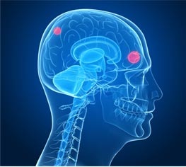
Metastasis is a word that simply means “spread”. So brain metastases are cancers that originated somewhere in the body outside the brain and have “spread” by the blood to lodge in the brain where the metastases (tumors that have spread) grow and damage the normal nearby brain. Any tumor can spread to the brain, but the most common tumors to do so are lung cancer, breast cancer, colorectal cancer, pancreatic cancer, kidney cancer, bladder cancer and melanoma. Lung cancer, the leading cause of cancer deaths in the United States, and breast cancer, the most common cancer in women, are responsible for more than half of the cases of brain metastases in the United States. Metastases to the brain may be single, meaning there is one tumor, or multiple meaning that there are two or more tumors that have spread to the brain. It is estimated that well more than 100,000 new cases of brain metastases occur in the United States each year.

Brain metastases are special because of the blood brain barrier. The blood brain barrier is unique, because it prevents many types of toxic chemical from reaching the brain, and this includes most types of chemotherapy. So any cancer cell that is able to slip through the blood brain barrier and into the brain itself is completely protected from the chemotherapy and may multiply to form a tumor which can damage the normal brain and cause symptoms. The symptoms caused by brain metastases depend on the area of the brain where the metastasis is located and what body functions that area of the brain controls. Common symptoms of brain metastases include headache from brain swelling, dizziness, weakness of a specific body part, poor coordination, problems with thinking or memory and possibly seizures.

Depending on several factors including the age and health of the patient, the type of cancer that has spread to the brain, the size and location(s) of the tumors within the brain, and the number of tumors possible treatments include surgery and post-operative radiation, or radiation alone either with whole brain radiation or with stereotactic radiosurgery. Stereotactic radiosurgery involves focusing high doses of radiation exactly on the metastases. Most commonly, stereotactic radiosurgery is given as a single large dose of radiation, but sometimes this can be given with a few separate treatments. This has the advantage of sparing much of the normal brain tissue from the effects of radiation and providing the best chances of long term control of the metastases. Recent studies have demonstrated that selected patients with 3 or fewer brain metastases may have improved tumor control and survival compared to patients receiving conventional whole brain radiotherapy.

Each case of brain metastases is unique. The expected duration of treatment and any side-effects of treatment must be discussed in detail with the physician who will be delivering the treatments.

Primary Spinal Cord Tumors

Tumors that begin in the spinal cord (primary spinal cord tumors) are very rare and occur in about 1 per 100,000 people. Metastatic cancer (secondary cancer or cancer that has spread to the spine from elsewhere) is far more common. Because these primary spinal cord tumors are so rare, very few clinical trials have been performed to determine in a systematic manner the best treatment of these tumors. As such, doctors use a “best common sense” approach and custom tailor the treatment to each patient as best suits his or her needs.

In the broadest of terms, primary spinal cord tumors can be divided into 2 groups. Tumors arising from the covering of the spinal cord or from nerves coming off the spinal cord. These include meningiomas, schwannomas, and neurofibromas. The other type of primary spinal cord tumor arises from cells making up the spinal cord itself. These include astrocytomas, ependymomas, hemangioblastomas and lipomas. There are other types of primary spinal cord tumors, but these are the most common of these overall very rare tumors.

The spinal cord is best thought of as an interstate highway that nerve impulses (traffic) uses to connect the brain and the rest of the body. This is a two-way street. Impulses come from the brain to the body to control muscle action, and from the body to the brain to carry touch sensations. Primary spinal cord tumors can affect either lane of traffic, and often affects both. Initial symptoms of primary spinal cord tumors reflect interference with this traffic. This includes interference with the brains ability to cause muscles to move resulting in muscle weakness and problems with walking and balance or possibly loss of control of the bowel or bladder. Likewise, interference with touch sensations traveling to the brain usually results in numbness but can also result in unusual sensations or even pain.

Because of the risk of damaging the spine and disrupting this essential traffic, surgery is difficult at best, and carries a high risk of damage to the flow of nerve impulses. Because of this risk of severe collateral damage, surgery, and sometimes even biopsy, cannot be safely performed. In those cases, the treatment of choice is radiation. In certain cases, chemotherapy may also be used along with radiation.

Unfortunately, the spinal cord has a maximum safe dose of radiation that can be given as treatment. Often, this safe dose of radiation, about 25 treatments, is not enough to gain long term control of the tumor. Eventually, tumors that have received maximum safe doses of radiation may begin to grow again. In these cases, additional radiation is not possible without high risk of damage to the spinal cord. Chemotherapy is an option to slow down tumor growth in cases where maximum safe doses of radiation have been given.

Bone Metastasis (Secondary Bone Tumors)

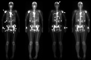
Bone metastases, also known as secondary tumors of bone, are cancerous tumors that have started elsewhere in the body and have traveled via the blood stream to become lodged in the bone where they grow and produce damage to the bone. This is very different from “bone cancer” which is a cancerous tumor which comes from the cells making up the bone. Bone metastasis is very common. The usual cancers which spread to bone include cancers which start in the breast, prostate, lung, pancreas, colon, rectum, kidney and others.

The main symptom of bone metastasis is usually pain in the part of the bone where the cancer has spread. When cancer spreads to a bone, it may weaken the bone and can cause it to break. Radiation is often used to help relieve pain from bone metastasis and also to prevent further damage to the bone allowing the bone to heal and reducing the risk of bone fracture. This risk of fracture is especially high in weight bearing bones such as the femur and tibia, but is also true of any bone which bears weight. This includes the vertebrae of the spine and the bones of the arms and pelvis. When bone damage is minor and is limited to a few spots, external radiation is usually given. When bone damage is minor and there are many spots, then various radioactive drugs can be given intravenously. When bone damage is severe and the risk of fracture is high, a surgical procedure, such as placement of metal rods or injection of a concrete like substance is usually done prior to external radiation to stabilize the weak bone and reduce the risk of fracture.

Overall, radiation for bone metastasis is a treatment commonly performed to reduce pain or to reduce the risk of fracture of the bone involved by cancer metastasis.